Do you scour the internet for 'liver tissue stained with massons trichrome biology essay'? You can find your answers here.
Table of contents
- Liver tissue stained with massons trichrome biology essay in 2021
- Diagnostic application of masson's trichrome
- Masson trichrome stain principle
- Mass on liver
- Masson trichrome stain heart
- Liver tissue stained with massons trichrome biology essay 06
- Liver tissue stained with massons trichrome biology essay 07
- Liver tissue stained with massons trichrome biology essay 08
Liver tissue stained with massons trichrome biology essay in 2021
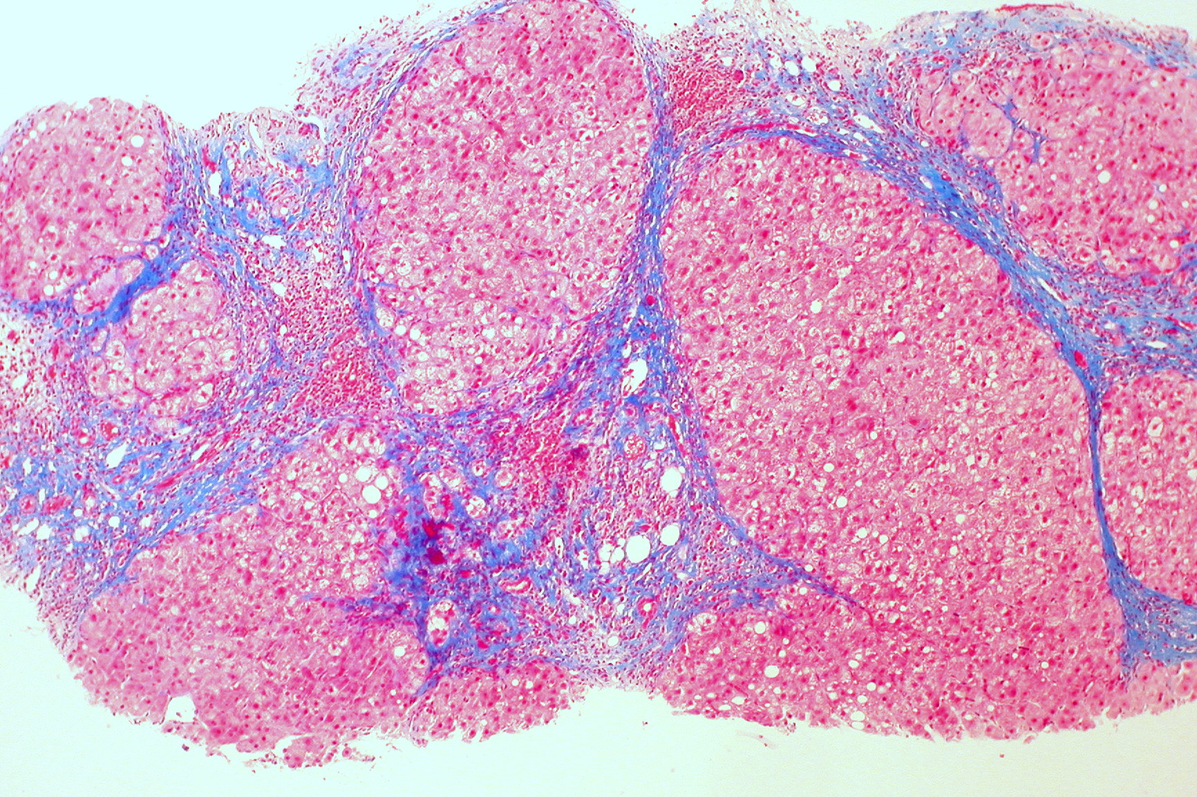
 This picture illustrates liver tissue stained with massons trichrome biology essay.
Diagnostic application of masson's trichrome
 This picture illustrates Diagnostic application of masson's trichrome.
Masson trichrome stain principle
 This image demonstrates Masson trichrome stain principle.
Mass on liver
 This image illustrates Mass on liver.
Masson trichrome stain heart
 This picture demonstrates Masson trichrome stain heart.
Liver tissue stained with massons trichrome biology essay 06
 This picture representes Liver tissue stained with massons trichrome biology essay 06.
Liver tissue stained with massons trichrome biology essay 07
 This image shows Liver tissue stained with massons trichrome biology essay 07.
Liver tissue stained with massons trichrome biology essay 08
 This image illustrates Liver tissue stained with massons trichrome biology essay 08.
How is trichrome staining used to detect collagen?
To stain collagen. To stain keratin. To stain fibrin. To stain muscle fibers. By use of the three stains, Masson’s Trichrome staining technique is used for the detection of collagen fibers in tissues such as the skin, heart, muscles.
How is trichrome staining used in liver biopsies?
Masson ‘s Trichrome discoloration is a common histology particular discoloration for liver and kidney biopsies. Liver cirrhosis is good demonstrated with this discoloration. In Masson ‘s Trichrome staining method, three dyes are used selectively for staining collagen fibers, musculuss, red blood cells and fibrin.
How is Masson trichrome used to diagnose liver cirrhosis?
Masson Trichrome Stain: Liver Cirrhosis Detection. Presently, therapies emphasizes on decreasing the supply of amyloid fibril precursor protein, while substituting and supporting the task of the involved organs. Liver cirrhosis and amyloidosis if not treated can lead to many complications.
What kind of stain is Masson's trichrome staining?
Masson’s Trichrome Staining is a histological staining method used for selectively stain collagen, collagen fibers, fibrin, muscles, and erythrocytes. It uses three stains for staining hence the term Trichrome. These are Weigert’s Hematoxylin, Biebrich scarlet-acid fuschin solution, and Aniline blue. To stain the collage fibers.
Last Update: Oct 2021